Healthcare Trends: Electrosurgery
The concept of using heat as a form of therapy and treatment to stop bleeding has been used for centuries, and was initially known as thermal cautery where tissues were burnt by thermal heat, including steam or hot metal, with the intention of destroying damaged or diseased tissue to prevent infections and reduce bleeding.
 Today, electrosurgery treatment is far more advanced and involves the controlled production of electrically induced heat through the passage of high frequency AC currents through biological tissue, allowing the current to cut or coagulate the tissue at the same time, minimising blood loss, shortening operating times and aiding faster patient recovery.
Temperatures over 45°C can cause the normal cell function to be inhibited and between 45°C and 60°C coagulation occurs causing the cell protein to solidify. Increasing the temperature further to 100°C produces desiccation and evaporation of the aqueous contents. Beyond 100°C carbonization occurs and the solid contents of the cells are reduced to carbon.
Polar Approach
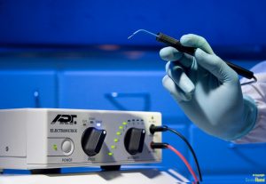
In electrosurgery, there are essentially two ways of delivering the required heat needed to undertake patient operations: monopolar and bipolar. The monopolar circuit requires electrical current to flow through the human body, while in the bipolar system the current flows from one tine to the other through the tissue held between forceps.
Monopolar electrosurgery is the most commonly used mode in surgery and used for cutting, fulguration and desiccation. It is usually represented by the Bovie pencil (small single probe), which is an active electrode located at the surgical site.
The electrical current flows from the active electrode through the patient’s body to the patient return electrode and then back to the generator. A high current density is produced at the tip of the probe which results in thermal heating and localised destruction whilst the return electrode, located on the patient’s body away from the surgical site has a large surface area and low current density. This is necessary to complete the circuit and prevent alternate burn sites as the high frequency AC current leaves the patient’s body.
In bipolar electrosurgery, the active and return electrodes are both located at the site of surgery, typically within the instrument tip – usually forceps. The current pathway is confined to the tissue grasped between the forcep tines, with one tine connected to one pole of the generator (active electrode) and the other to the opposite (return electrode).
Therefore no patient return electrode is required to complete the circuit and the patient’s body does not make up part of the electrosurgical circuit as only the intervening tissue between the tines contains the high frequency electrical current.
Due to the small amount of tissue held in the instrument much lower voltages are required and the thermal energy produced is evenly dispersed between the two electrodes, coagulating the tissue with minimal thermal damage to surrounding tissue. It is used for desiccation without sparking, avoiding damage to adjacent tissue caused by the arc and spraying of high frequency current and is used in delicate highly conductive tissue.
ESUs can be programmed to function in several modes with distinct tissue characteristics and output can be varied in two ways: the voltage can be altered to drive more or less current through the tissues, or the waveform can be modified which influences the tissue effect. The tissue effect associated with the different electrosurgical current waveforms is dependent on the size and shape of the electrode and the output mode of the generator.
There are three types of current waveforms: cutting, coagulation, and blended currents. Cutting currents use an uninterrupted sinusoidal waveform with high average power, high current density and a crest factor (CF, ratio between peak and RMS voltage) of 1.4. The use of electric sparks allows for precise cutting and focused heat which minimises widespread thermal damage.
The electrode should be held slightly away from the tissue to create a spark gap and discharge arc at specific locations which produces a sudden and localised heating effect over a short period of time which causes extreme heating and vaporisation of intracellular fluid that bursts cells. A fine, clean incision is created through the biological tissue with minimal coagulation (haemostasis) or extensive thermal damage and the continuous current does not allow for tissue cooling.
Coagulation currents are characterised by high voltage intermittent bursts of dampened sine waves which drive the current through the tissues and relatively low current which reduces the duty cycle to 6%. Coagulation currents typically have a CF of around 10 and involve electrical sparking over a wide area, creating less heat and resulting in evaporation and relatively slow dehydration which seals blood vessels while keeping cells intact.
A blended current is a modification of the duty cycle and operates at voltages between those of cutting and coagulation with a CF usually in the range of 3 to 10. They allow for tissue division while maintaining a variable degree of haemostasis which is defined by the off period.
Although the total energy remains the same, the ratio of voltage and current is adjusted to increase haemostasis – achieved by interrupting the current and increasing the voltage, to deliver a waveform in intermittent bursts. Modifications and reductions to the duty cycle through progressive blends produce less heat and as the interval between bursts progressively increases, greater coagulation is produced.
The rate at which heat is produced is the dominant factor and only variable in determining whether a waveform vaporises or coagulates biological tissue. Surgeons have the option to combine the cut and coagulate currents to produce different tissue effects. Coagulation can be performed with the cutting current by using the electrode in direct contact with the tissue and this requires less voltage than the coagulation waveform. However power settings may need to be adjusted and electrode size varied to achieve the desired surgical effect [1].
Testing generators
The original ESUs were ground referenced where the electrical current passed through the patient’s body and returned to ground. The grounding is intended to occur via the patient return electrode which is usually situated on the thigh of the patient and away from the surgical site. However, electrical currents seek to travel down the pathway of least resistance and therefore current can travel through any conductive grounding object that is in contact with the patient as a method of ground return, such as ECG electrodes or tables and operating staff. This increases the possibility of creating alternate site burns on the patient at alternative grounding sites where the high frequency current has exited the patient.
Although many manufacturers no longer rely on ground referenced ESUs due to the high risk of skin burns associated with alternative grounding more modern units must still be tested regularly to ensure that they are operating correctly and safe to use – for both the operator and patient. But before proceeding further it is important to understand the operation of the generator or device under test (DUT). The output energy of electrosurgical generators can lead to burn injuries. Always ensure that the tests are conducted by a suitably trained individual and limit the amount of accessible conductive parts that become live with high frequency electrical current.
To maximise safety, there have been a number of accessories developed from companies to automate the testing and reduce the need for manual interaction during testing and while the output of electrical surgical generators are active. These include the new Rigel Medical Uni-Therm electrosurgical analyser, one of the quickest and easiest ways to test electrosurgical generators using single instrument, combining the test functions to verify the CQM, the high frequency leakage and the output power. By providing built-in automation and data storage, the Uni-Therm can be deployed for in-service applications as well as at the end of demanding production lines or in-test laboratories.
To maximise the effectiveness of the surgical procedure and to reduce the risk of injury during electrosurgical procedures, the patient plate must cover an optimum amount of skin surface area (quantity) and be high in conductivity (quality) where the energy exits the patient. This is monitored by the electrosurgical device through impedance measurement (CQM) between the two (split) or more conductive pads within the patient return plate. When extreme variations or very high/low impedance appears, the CQM will lead to an audible and/ or visual alarm and can lead to deactivation of the output energy to prevent potential patient injury.
The Rigel Uni-Therm facilitates accurate CQM function simulation, allowing automatic and manual increase or decrease of electrical resistance values in 1Ω resolution, and enabling the testing of modern contact quality monitoring systems that are triggered by relative changes in resistance.
Unlike conventional analysers, the Rigel Uni-Therm utilises a motor driven potentiometer which can simulate resistance variations between 0–475Ω to within 1Ω resolution. This allows the user to trigger the CQM system by simulating fault conditions including high or low impedance values or a large variation in impedance, for example a change of 10%. Impedance can also be controlled manually by utilising the rotary encoder on the front panel.
Design criteria of electrosurgical generators (IEC 60601-2-2) require the manufacture to limit the amount of capacitive leakage of the high frequency current. At frequencies exceeding 400 kHz, the electrical current has a tendency to stray, leading to a decrease in functionality and possible injury to the patient.
Capacitive coupling might occur between the test leads during the setup. This is the reason why IEC 60601-2-2 stipulates specific layout of test leads and test loads to ensure the capacitive coupling is limited and controlled in a laboratory environment, these tests are referred to as the long lead tests. A more practical approach is to ensure the test leads are as short as possible and do not cross over, to limit the influence of capacitive coupling.
Breakdown of insulation in the surgery leads as a result of high voltages (peak-to-peak up to 10kV) is also a consideration when testing the electrosurgical generator. This can be verified by including the surgical leads as part of the test setup. The HF leakage test measures the HF leakage current in various test configurations and compares the result to a user set pass/fail value using the rotary encoder to navigate the screens. The Uni-Therm simplifies the complex test configurations of high frequency leakage current measurement, as required by IEC 60601-2-2, by providing detailed instructional diagrams for each high frequency leakage test set-up on its colour display.
The ability to measure currents of up to eight Ampere RMS is important and can be achieved through the use of a customised current transformer, capable of accurately measuring high currents when calibrating electrosurgical generators with high current vessel sealing treatment functions.
The power measurement options in the Uni-Therm include measuring output power and current during a single load value (continuous), the output power and current under a changing load condition (graph) and the output and current during short circuit testing or when using a specific external load resistor during development (external load). The large colour display provides a clear and detailed interpretation of output power whilst cut and coag foot paddle control automates the process; making this a fast, effective and safe test procedure.
Graphical representations of power distribution curves can be easily switched to numerical data at the touch of a button without the use of a PC and the Rigel Uni-Therm will control the device under test (DUT) by using the internal footswitch controller with a footswitch adapter leading from the footswitch connector on the ESU to the cut and coag sockets on the front of the Uni-Therm.
The whole test procedure for testing the electrosurgical generator can be programmed into the Rigel Uni-Therm. The cloning features make sharing of test configurations between different Uni-Therms simple, so it easier and faster to configure and update your test instrument. Each test step can be set up with simple user instructions for DUT settings such as mono or bipolar, energy settings and waveform selection. The CUT and COAG footswitch controls on the Uni-Therm can be used to control the electrosurgical generator. This can reduce the over-all test time and increase user safety.
Understanding requirements
There’s no question that the use of electrosurgical generators has led to more effective surgical treatments and improved patient safety through greater control and management of complications during surgery. Nevertheless, the use of ESUs is not without risk and remains one of the more hazardous practises in operating theatres.
Regular performance and safety tests must be seen as an essential part of any in-service maintenance programme and can lead to further improvement of patient safety by ensuring the safety features of each generator is intact, and that the performance accuracy is guaranteed. When considering the purchase of electrosurgical analysers, ensure that you understand the manufacturer’s requirements and the technical capability of your install base.
For instance, when calibrating electrosurgical generators with high current vessel sealing technology, look for test equipment that can measure both short circuit currents as well as currents over 5A RMS. Testers like the Rigel Uni-Therm are versatile and compact yet offer safer, faster and more accurate testing of electrosurgical generators, ultimately enabling the end-user to comply with and satisfy effectively both international and manufacturers’ specific test requirements.


